What is Raman Spectroscopy?
Raman spectroscopy is an analytical tool concerned with radiation scattering from a sample such that the structure, properties and environment can be studied based on vibrational transitions within the material. This technique is based on the Raman effect (inelastic scattering of light) discovered in 1928 by Indian Physicist Chandrashekhara Venkata Raman. Raman found that sunlight interacted with molecules to produce a characteristic pattern when a small fraction of the incident radiation is scattered by the molecule and that it differed in wavelength from that of the incident beam. It was also noted that the shifts in the wavelength depended on the chemical structure of the molecules responsible for the scattering.
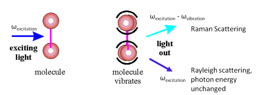
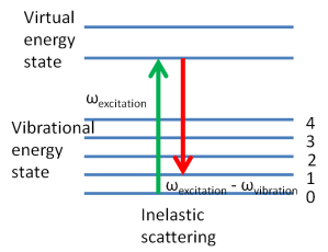
So, in the collection of a Raman spectrum of a material monochromatic light, usually a laser hits a molecule as seen above. The molecule experiences a momentary distortion of the electrons distributed around a bond by the incoming photon from the laser which creates an induced dipole moment in the molecule followed by re-emission when it relaxes. When the molecule relaxes the frequency of some of the photons is changed (Raman scattering) and do not return to their original state but is shifted to a lower energy state as seen below. The difference between the ground state and the new energy state is the Raman shift.
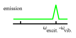
The different vibrational modes associated with the groups of the structure will give different Raman shifts that can then be plotted to give a fingerprint of the material of interest and this is what we know as the Raman spectrum.
Types of Raman Spectrometers
A Raman spectrometer is composed of a light source, momochromator, sample holder and detector, the Raman spectrometer can be one of two designs, dispersive or Fourier Transform. They differ in laser sources and the way by which Raman scattering is detected and analysed.
Advantages of Raman Spectroscopy
- Water does not cause interference as in FTIR spectroscopy and as such aqueous solutions can be tested and samples do not have to be dry.
- Glass or quartz cells can be used as sample holders
- No sample preparation is usually required
- Significantly there are several advanced techniques which allows Raman spectroscopy to be applicable to many areas. These advanced techniques include:
- Surface enhanced Raman Spectroscopy (SERS)
- Resonance Raman Spectroscopy (RRS)
- Confocal Raman microscopy (CRM)
Advanced Techniques
Surface enhanced Raman Spectroscopy (SERS) involves obtaining spectra in the usual way but is done on samples that have been adsorbed on the surface of colloidal metal particles or on a roughened surface. This technique has higher sensitivity as it results in stronger Raman signal, the signal can be enhanced about 103 to 106 times greater and allows for detection of single molecules. Many applications—biochemistry, chemical manufacturing, environmental detection, forensics.
Resonance Raman Spectroscopy
During normal Raman spectroscopy any wavelength laser can be used for excitation in order to measure the Raman scattering. In resonance Raman the excitation wavelength is selected to overlap with the electronic transition. This overlap can result in increasing scattering intensities by factors of 102-106, this allows for very low detection limits. Unfortunately, fluorescence backgrounds can be much higher than in normal fluorescence and can be more problematic depending on the sample.
Confocal Raman microscopy
In this setup a Raman spectrometer is combined with an optical microscope. The excitation laser beam is focused on the sample through the microscope objective where a micro-spot is created. The backscattered beam from the sample is re-focused onto a pinhole aperture that acts as a spatial filter where the CCD camera collects only Raman signal as it filters interference from Rayleigh scattering and fluorescence unlike in regular Raman where this interference is much higher.
Raman microscopy allows for analysis of individual particles and characterization of sample features with dimensions down to 0.5μm. Raman mapping of sample features to show distribution of components, with spatial resolution down to 0.5μm is also possible. Most significantly the addition of confocal optics gives depth spatial resolution allowing for multilayered samples to be analysed.
The use of all of these techniques makes Raman spectroscopy very useful for the material scientist. Its capabilities in providing information about chemical structure and identity, intrinsic stress/strain and phase and polymorphism makes its applications very wide and varied. It has been found to be very useful in studying semiconductors and superconductors, carbon nanotubes, polymers, environmental materials, ceramics, molecules and molecular systems and archeological materials.
See part II for more details on Application in Material Science
References
- Das, R.S. & Agrawal, Y. 2011 ‘Raman spectroscopy: Recent advancements, techniques and applications’ Vibrational Spectroscopy, Vol. 57(2), pp.163–176. Available: Elsevier/ScienceDirect/10.1016/j.vibspec.2011.08.003 , [5 Nov 2011]
- Larkin, P. (2011) IR and Raman Spectroscopy inIR and Raman Spectroscopy Principles and Spectral Interpretation, Elsevier Inc.
- Jimenez-Sandoval. S 2000 ‘Micro-Raman Spectroscopy: a powerful technique for materials research’ Microelectronics Journal, Vol.31,pp.419-427. Available: Elsevier/ScienceDirect/PII S0026-2692 (00) 00011-2 , [20 September 2012]
- Ian Lewis and Howell Edwards (ed.) (2001) Handbook of Raman Spectroscopy, Mercel Dekker, Inc., New York


This article about Raman Spectroscopy is getting really interesting. I am impressed with the content and i will look forward for the next part of this. Great post!